Interesting Cases and Pictures
This is where I’ll upload interesting cases and good pictures I take throughout residency/my career.
Arlt’s Line (Trachoma)
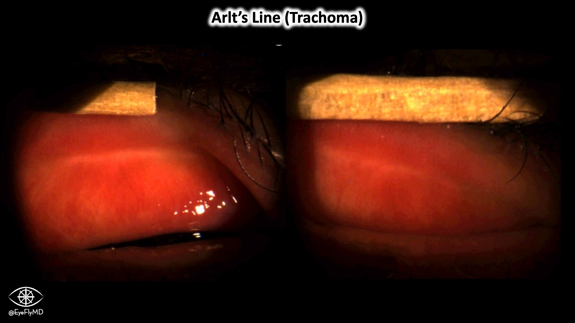
It’s not everyday you see an Arlt’s Line in the US! This band of scar tissue is indicative of trachoma (caused by Chlamydia trachomatis A, B, and C). Repeated, untreated infection can cause scarring, entropion, and blindness.
Traumatic Lens Dislocation
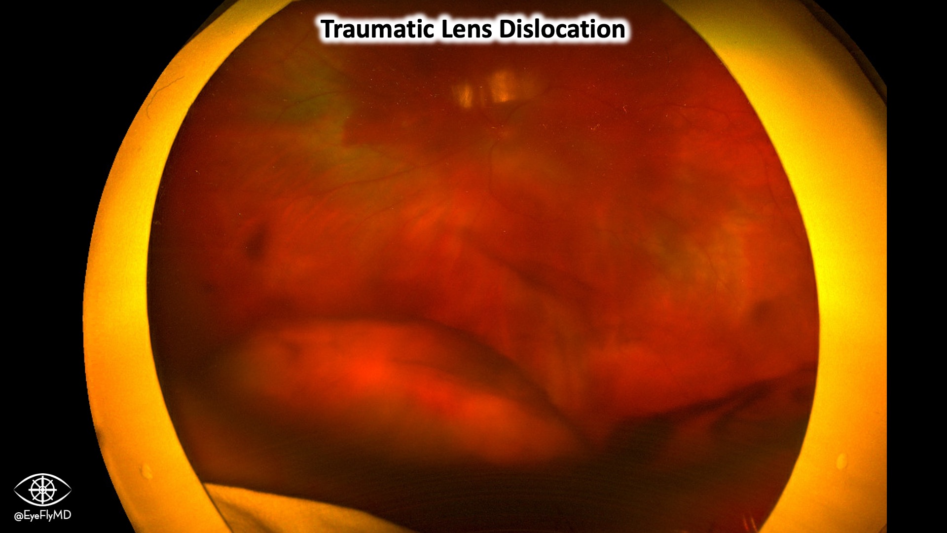
Appreciate the commotio, vitreous heme, and of course the crystalline lens residing in the posterior segment.
Epicapsular Star
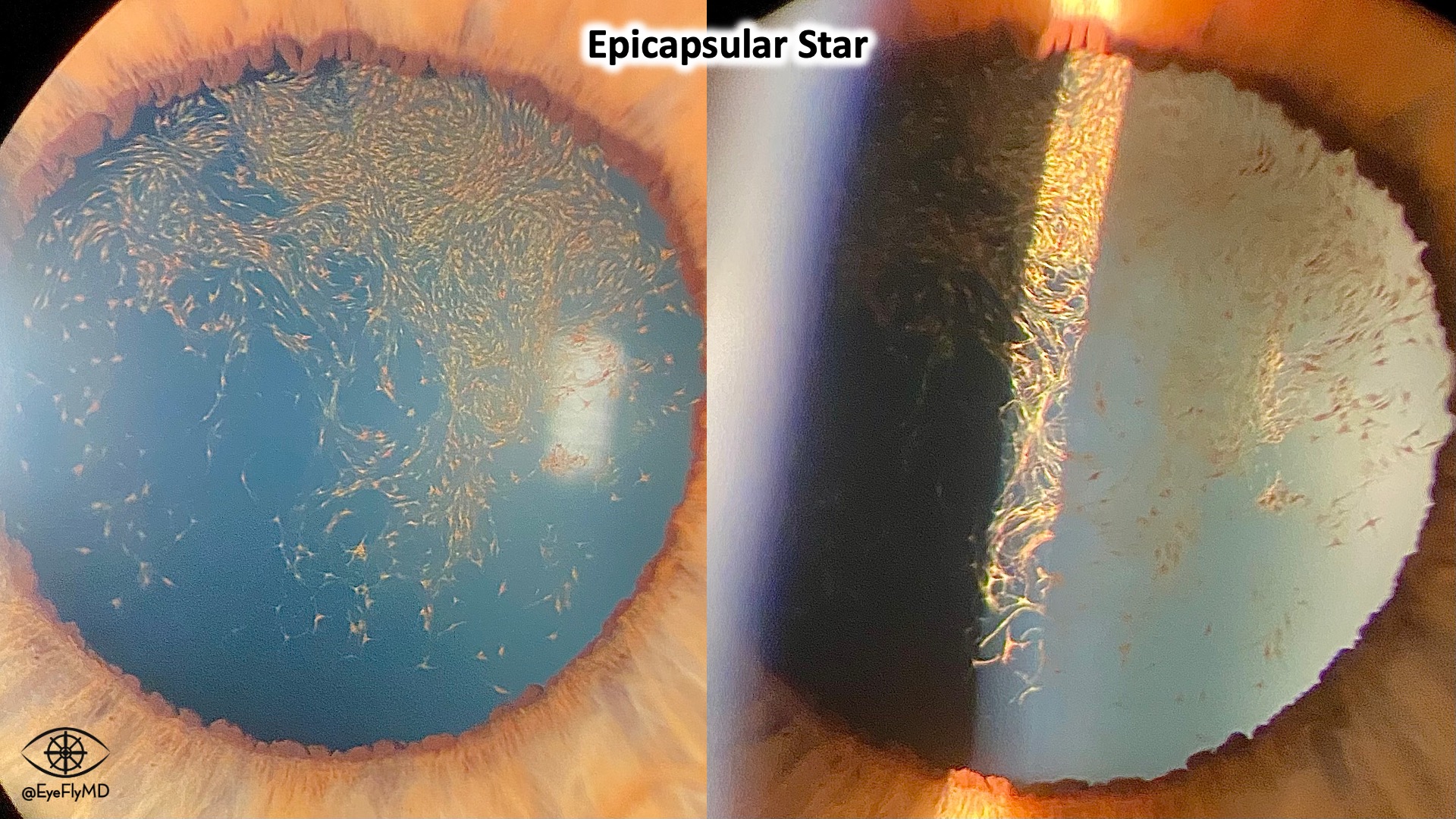
Epicapsular Star (aka chicken tracks) refers to the persistence of the anterior vascular portion of the tunica vasculosa lentis that derives from the long posterior ciliary arteries. This is a pretty extensive example.
Granulomatous KP in Arlt’s Triangle
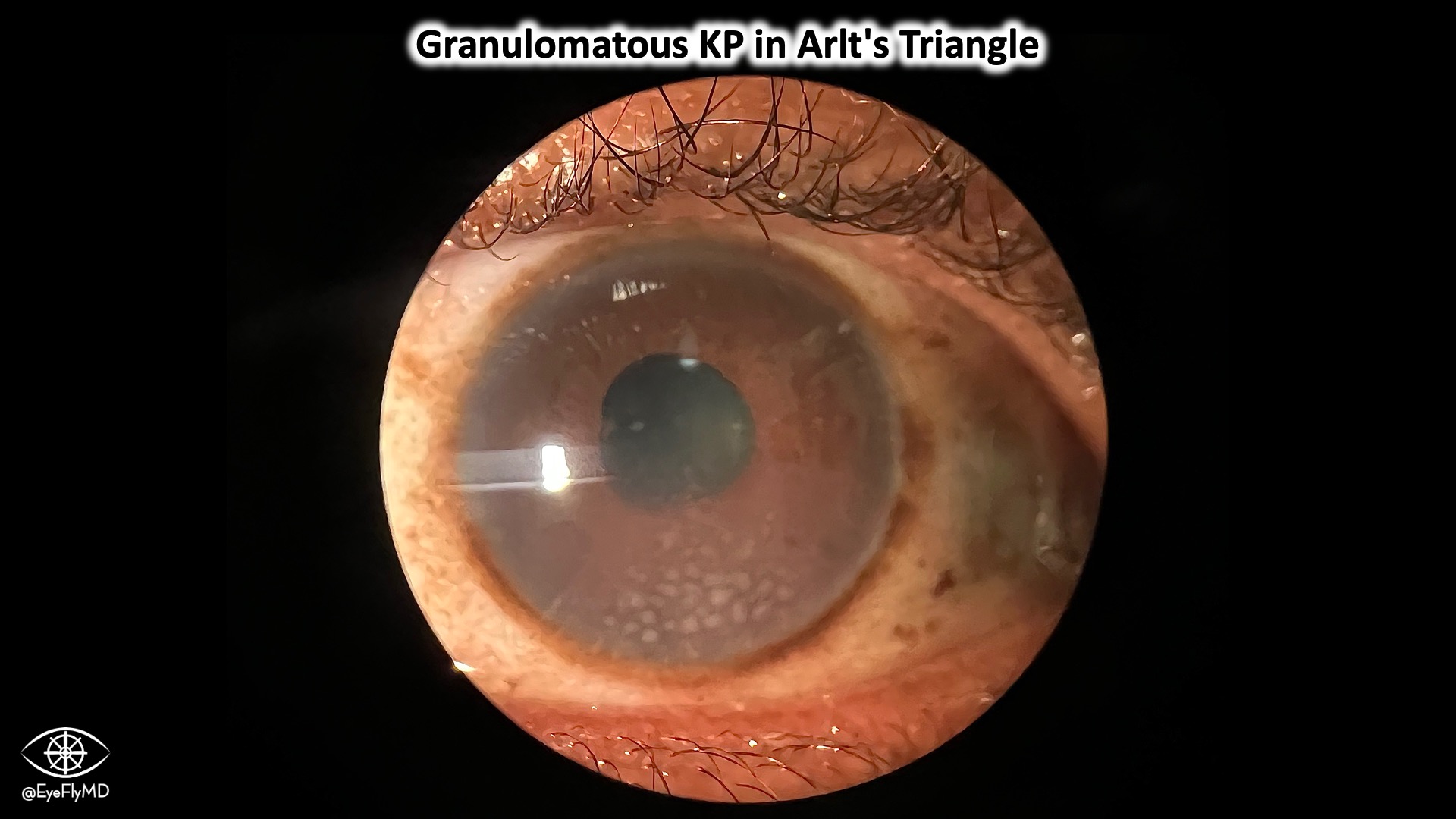
This is a good example of granulomatous KP perfectly within Arlt’s triangle. Arlt’s triangle refers to a triangular shape of cornea with the apex at the center of the cornea and base inferiorly. This should make you think of the big 3: Sarcoid, Syphilis, TB. Also remember: Lyme, VKH, SO, and toxocariasis. This pattern is due to gravity and aqueous fluid currents.
CRAO with Boxcarring of Vessels

side from the obvious cherry red spot you can appreciate the boxcarring in the vasculature and whitening of the macula.
Essential Iris Atrophy (ICE Syndrome)
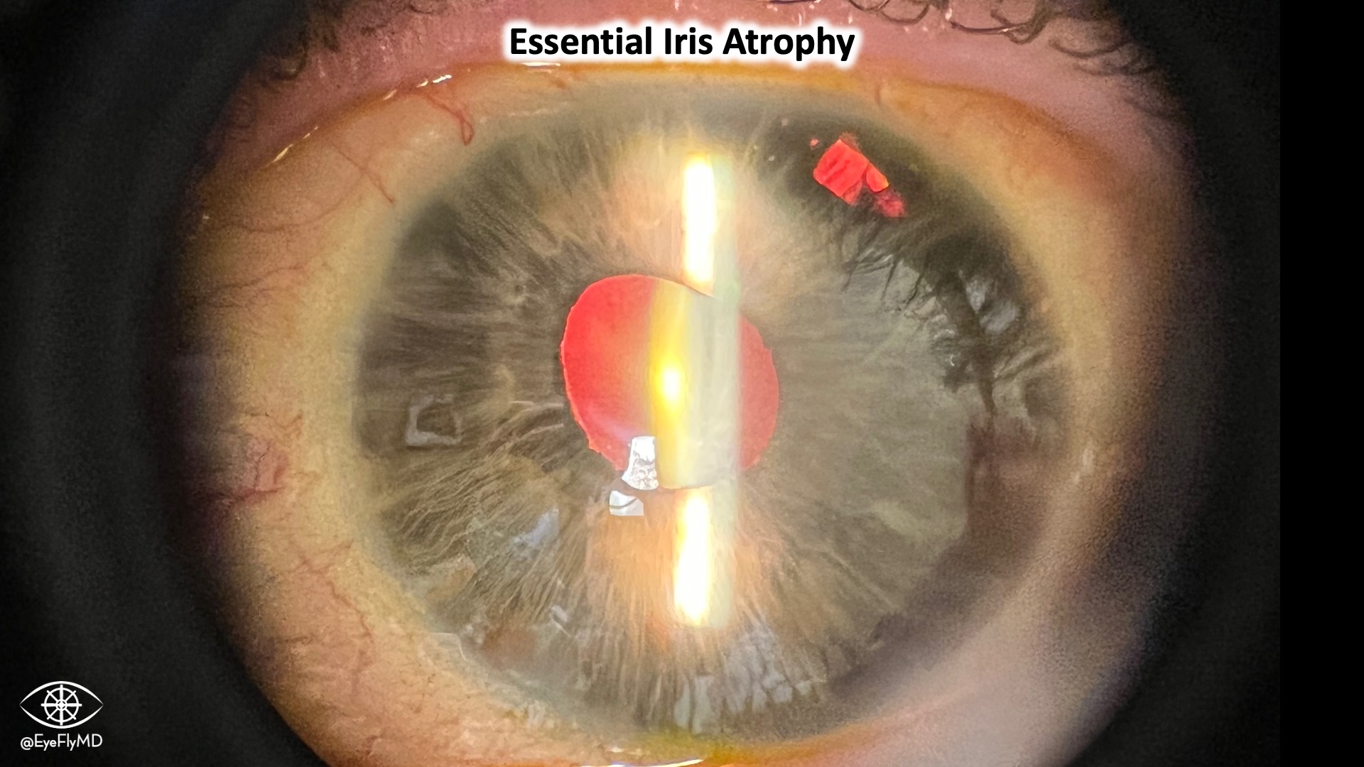
Good example of Iridicorneal Endothelial syndrome. Presented with vision change and IOP in the 60s! This is most consistent with the “Essential Iris Atrophy” variant.
Fuchs Heterochromic Iridocyclitis (FUS)
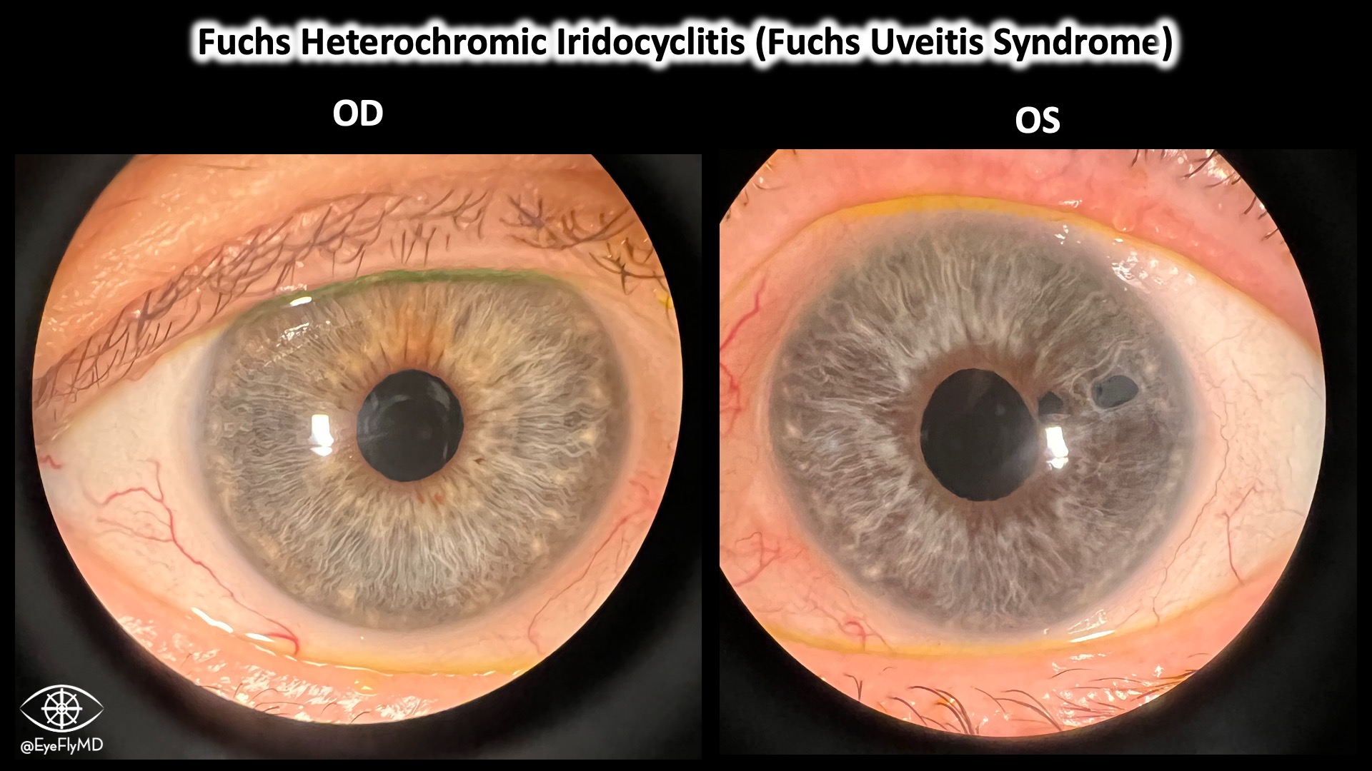
This is a unilateral, chronic glaucoma characterized by heterochromia iridis, cataract, and stellate KP. In dark eyes, the light eye indicates the affected side. In light eyes, it’s the darker one. This is also called Fuch’s Uveitis Syndrome.
Optic Disc Edema with Flame Hemorrhages
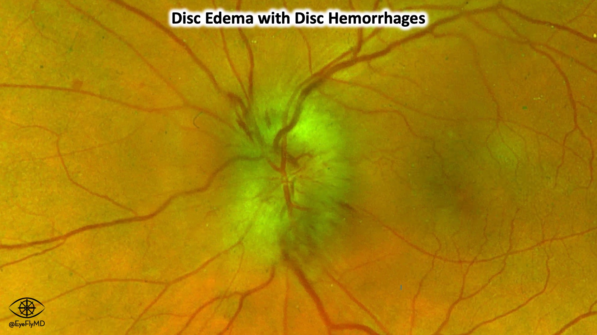
Drance/Disc/Flame hemorrhages, call them what you like but they are small hemes perpendicular to the optic disc and parallel to the RNFL. Here are disc hemes in with edema in a suspected case of NAION.
Vitreous Prolapse
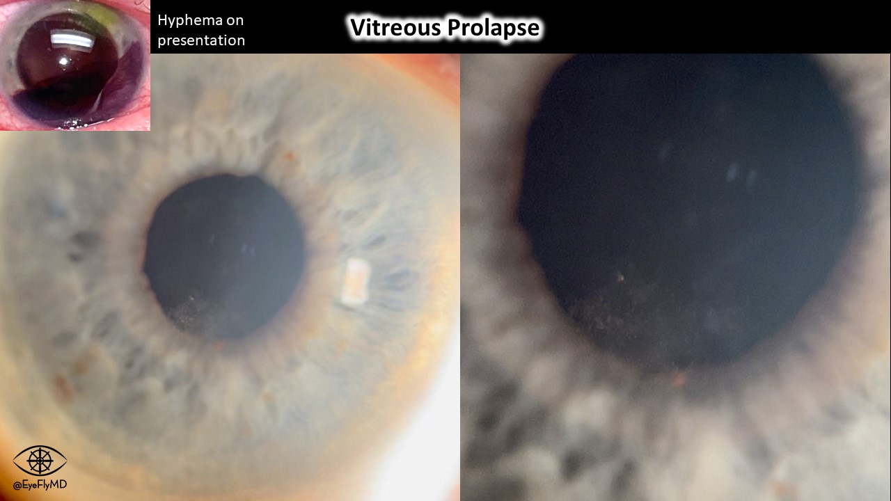
A case of weed-wacker propelled rock to the eye resulting in large traumatic hyphema and vitreous prolapse.
Keratoprosthesis
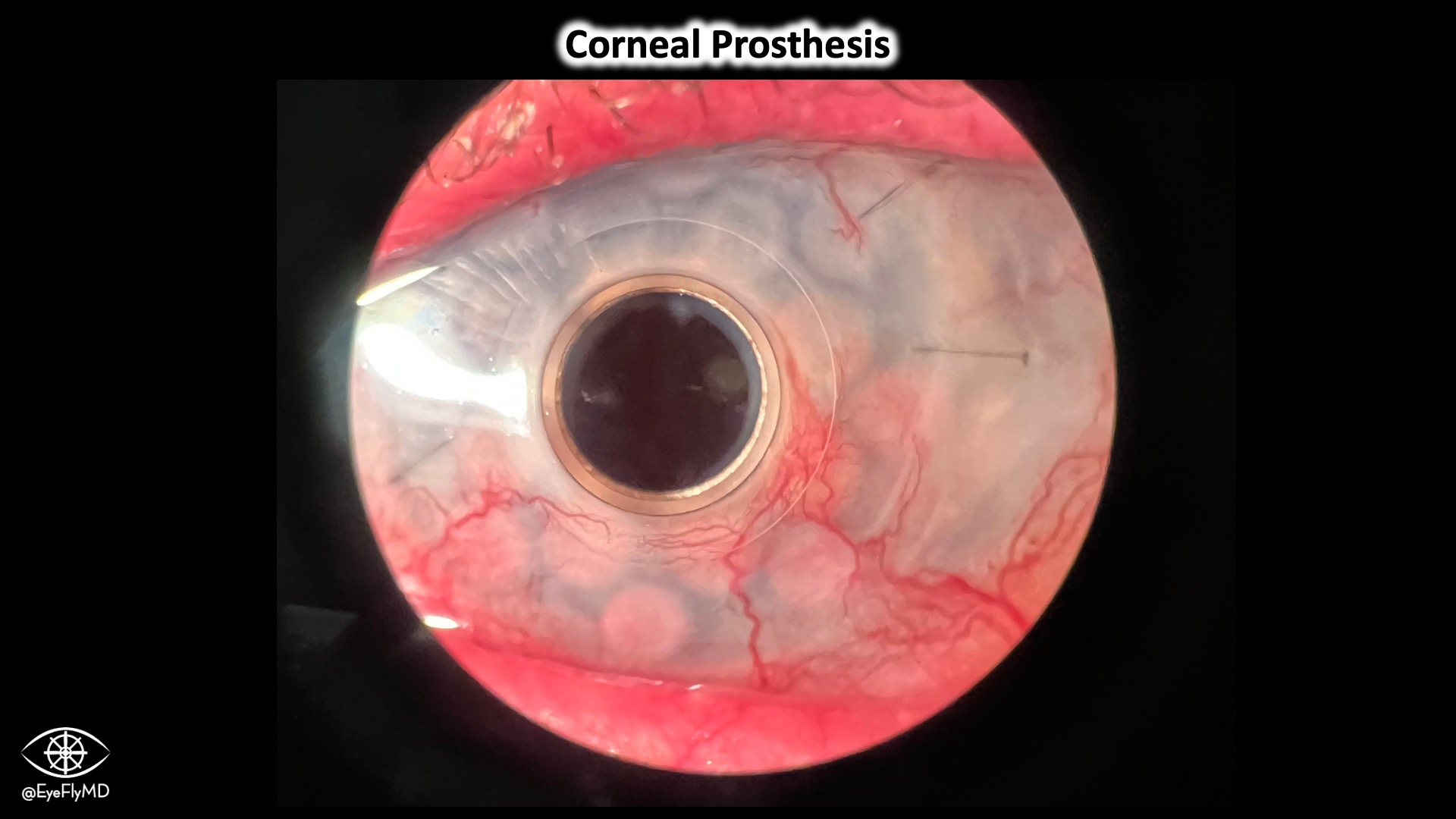
This is a TWELVE year old keratoprosthesis still going strong! These are reserved for multiple PKP failures.
Pseudoexfoliation
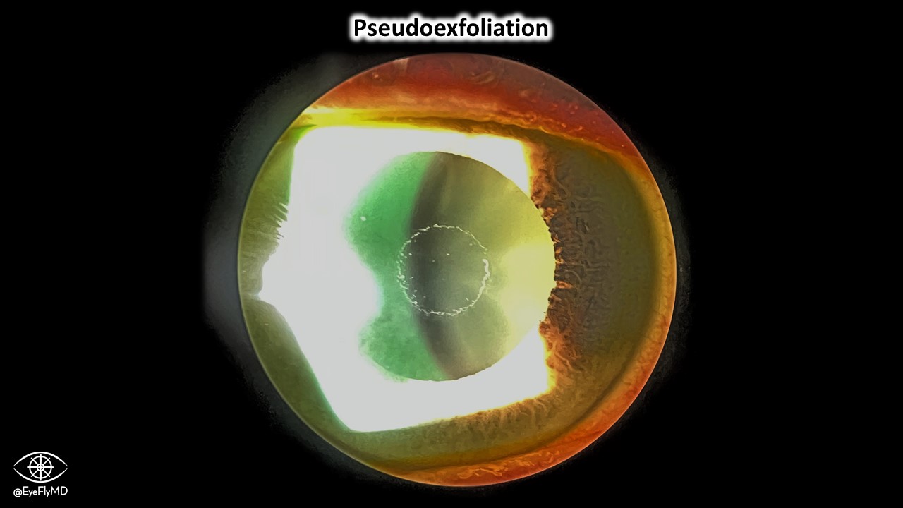
Excellent example of pseudoexfoliative material on the anterior lens capsule. Remember, “pseudo” because the anterior capsule is not actually delaminating because the patient is not a glass blower. This disease is characterized by flaky, fibriller, white material that is deposited throughout the body (not just the lens).
Torsional/Rotary Nystagmus
Great eample of torsional nystagmus likelye secondary to vestibular neuritis. It looks like SO myokymia but the fast phase is excyclotorsion OD and incyclotorsion OS.
Traumatic Cataract, Lens Subluxation

IOL Glistenings
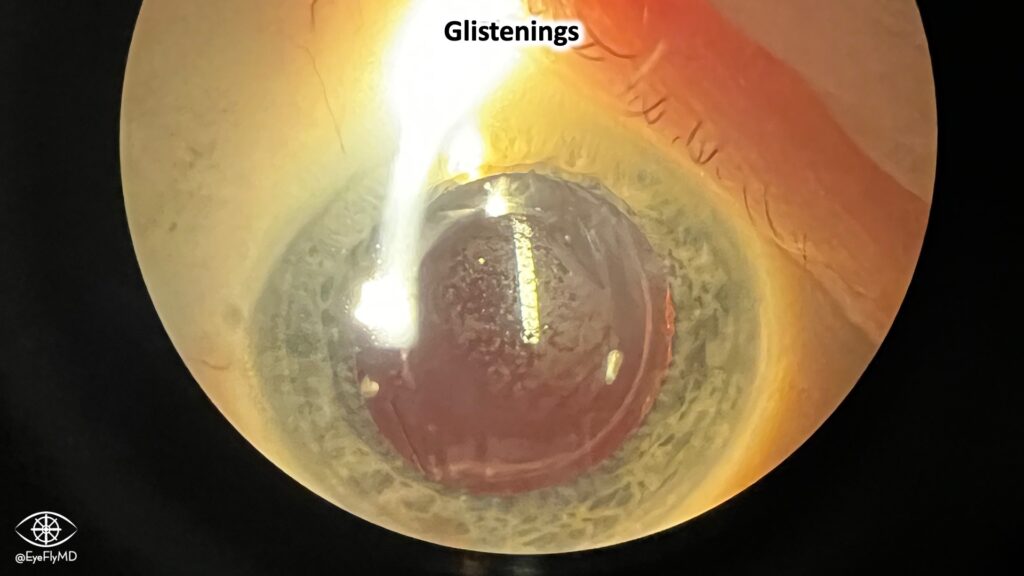
Glistenings are microvacuoles that form on hydrophobic IOLs. They’re not usually visually significant. These were common with the AcrySof platform but the updated Clareon material fixed this. EnVista is also a glistening-free IOL platform.
Vitreous Cyst
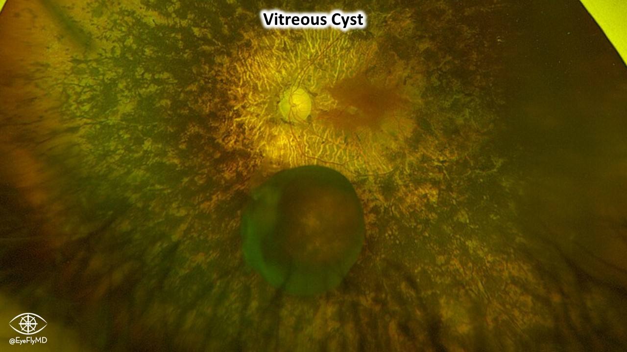
Patient with extensive retinitis pigmentosa and a decent sized vitreous cyst. Vitreous cysts are very rare and are a choristoma of the hyaloid system.
Tilted CT Lucia IOL
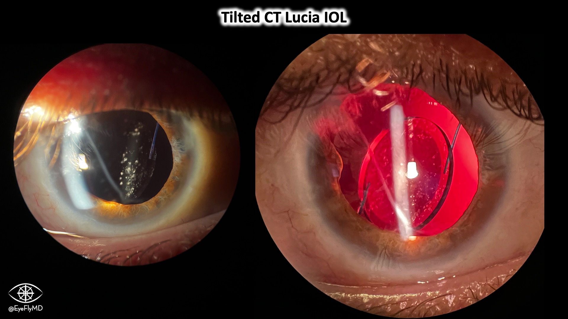
CT LUCIA is an IOL by Zeiss that uses special PVFD haptics that are more amenable to manipulation than PMMA haptics. There are case reports of these IOLs tilting post-operatively and this is a good, representative example.
Gore-Tex Sutured IOL

A cool feature of the MX60 (enVista) is the eyelets that allow or cases like this. The IOL was dislocated but subsequently sutured to the sclera using the eyelets.
Multifocal Choroiditis with Panuveitis (MCP)
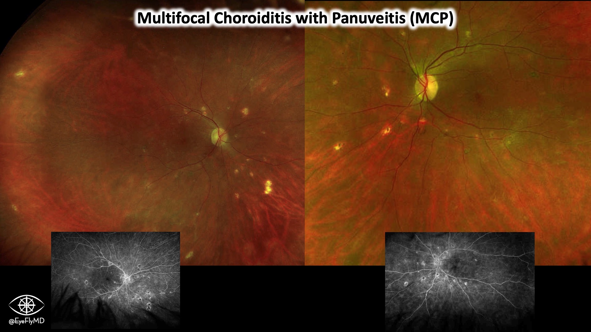
Anterior uveitis, vitritis, visual disturbances, and these fundus findings in a young myopic female is very consistent with Multifocal Choroiditis with Panuveitis. Aside from the vitritis, this looks a lot like POHS. In fact, an old term for this is “pseudo-POHS”.
Asteroid Hyalosis
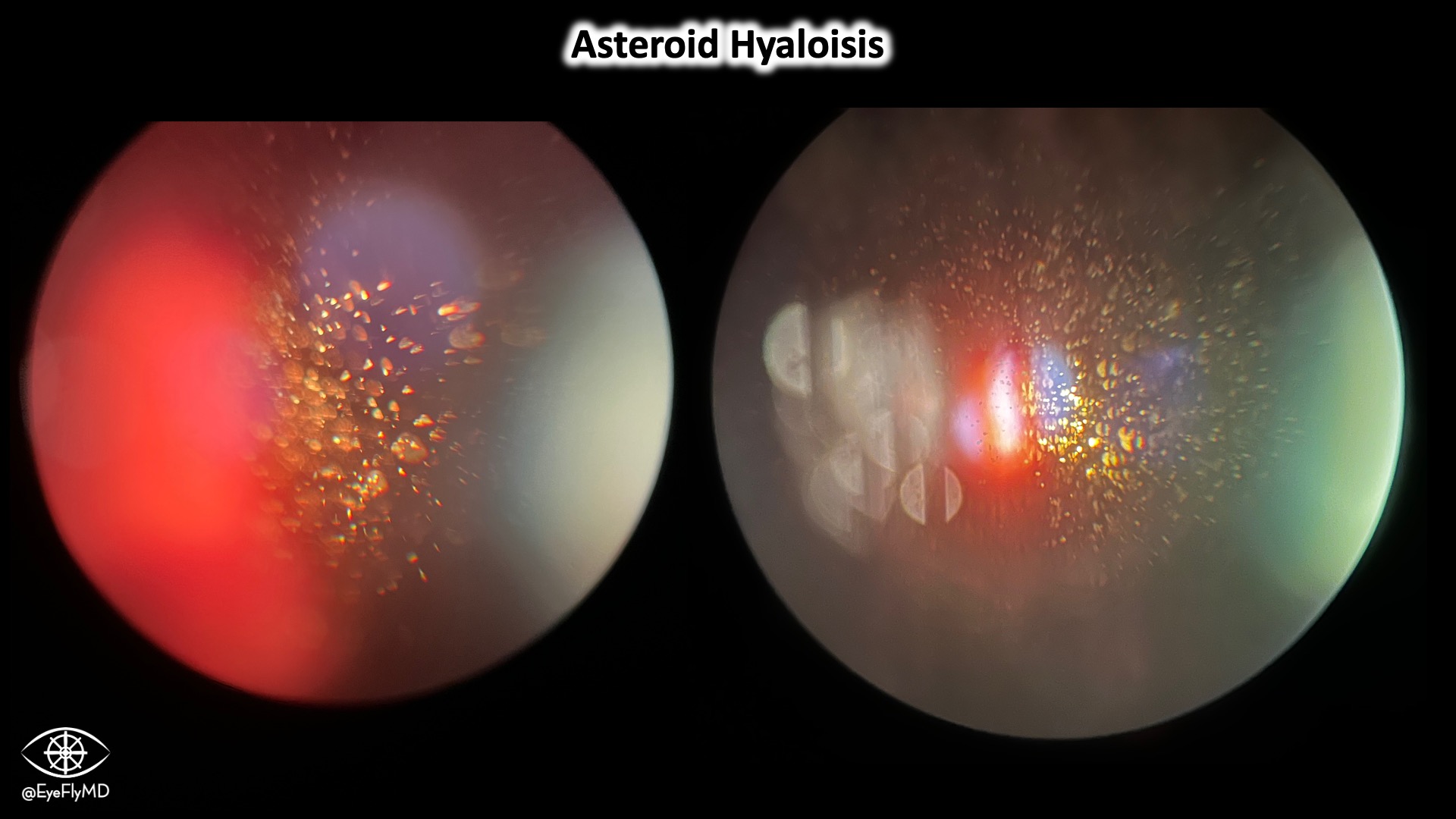
Asteroid hyalosis is a common condition characterized by deposits of lipids, calcium, and phosphorus suspended in the vitreous. It resembles an asteroid field. It’s associated with DM and differentiated from synchysis scintillans where the deposits float freely in the vitreous and settle.
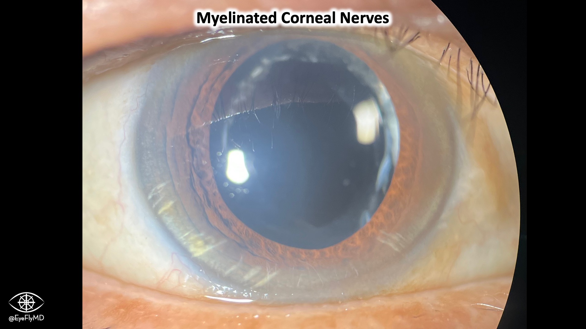
Myelinated Corneal Nerves
Capsular Bag Distension Syndrome
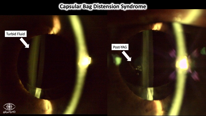
Capsular Bag Distension Syndrome involves the buildup of turbid fluid behind an IOL that can degrade visual acuity. A two shot YAG resolved this case.
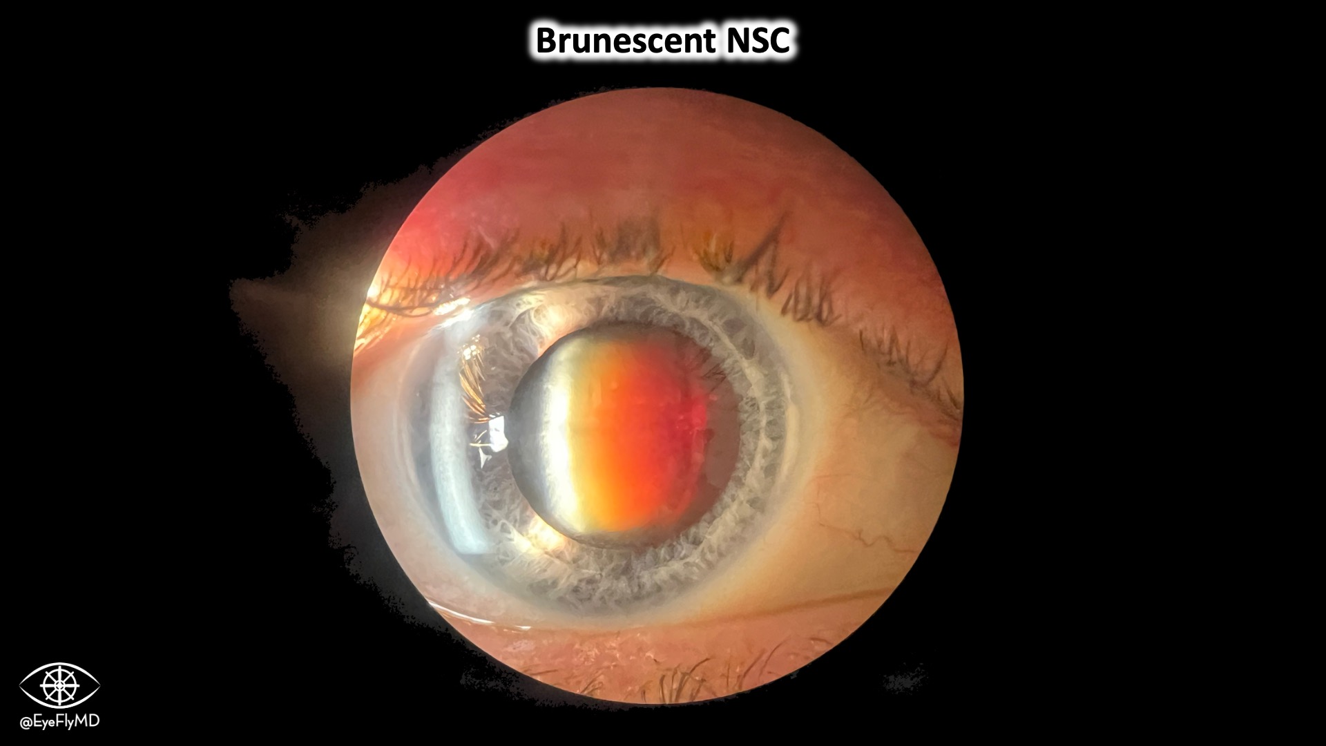
Brunescent MSC